
Prostate fusion biopsy made simple.
Prostate cancer is the only solid cancer still diagnosed through random sampling. Get accurate and meaningful results faster with a targeted approach.
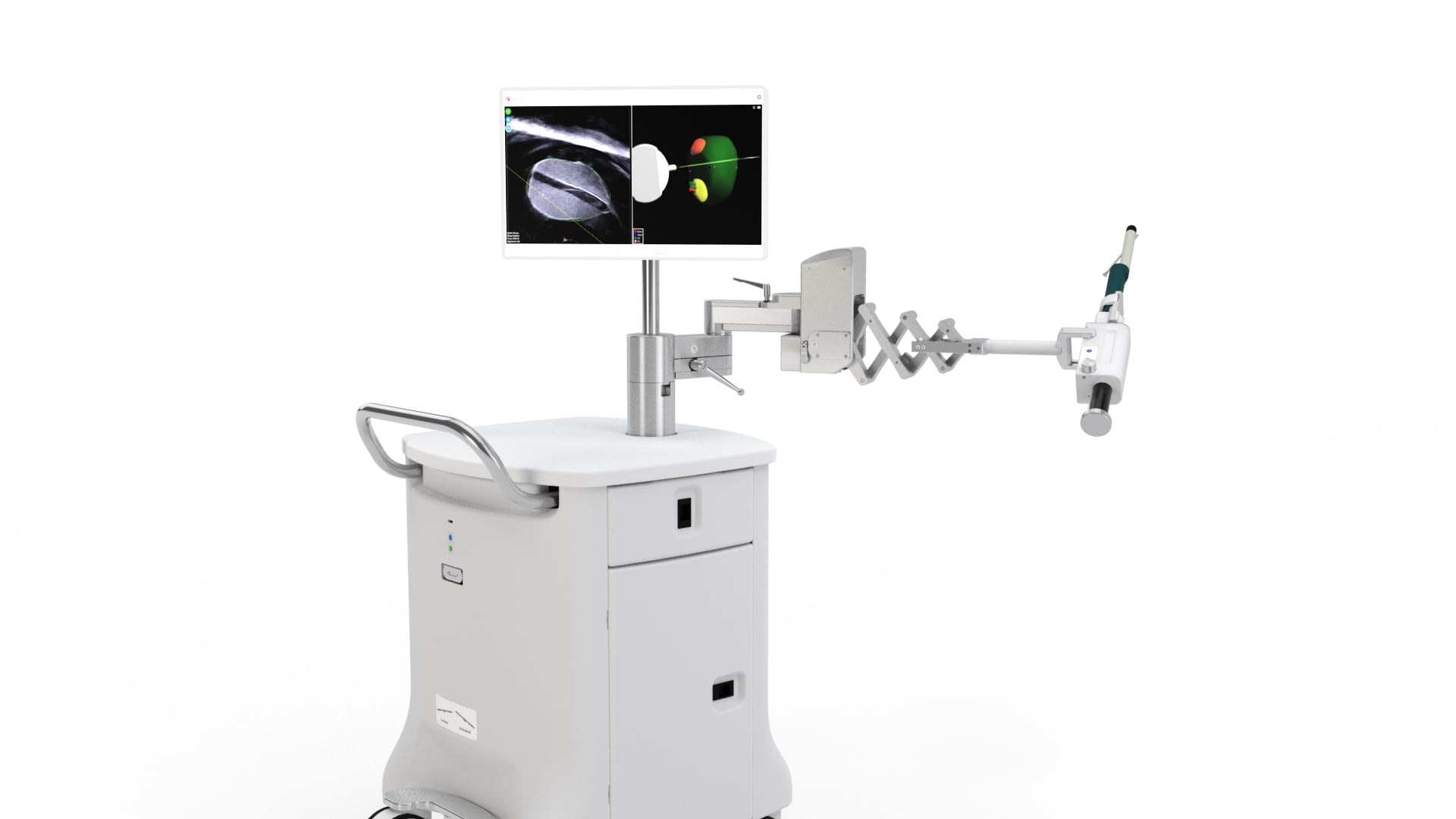
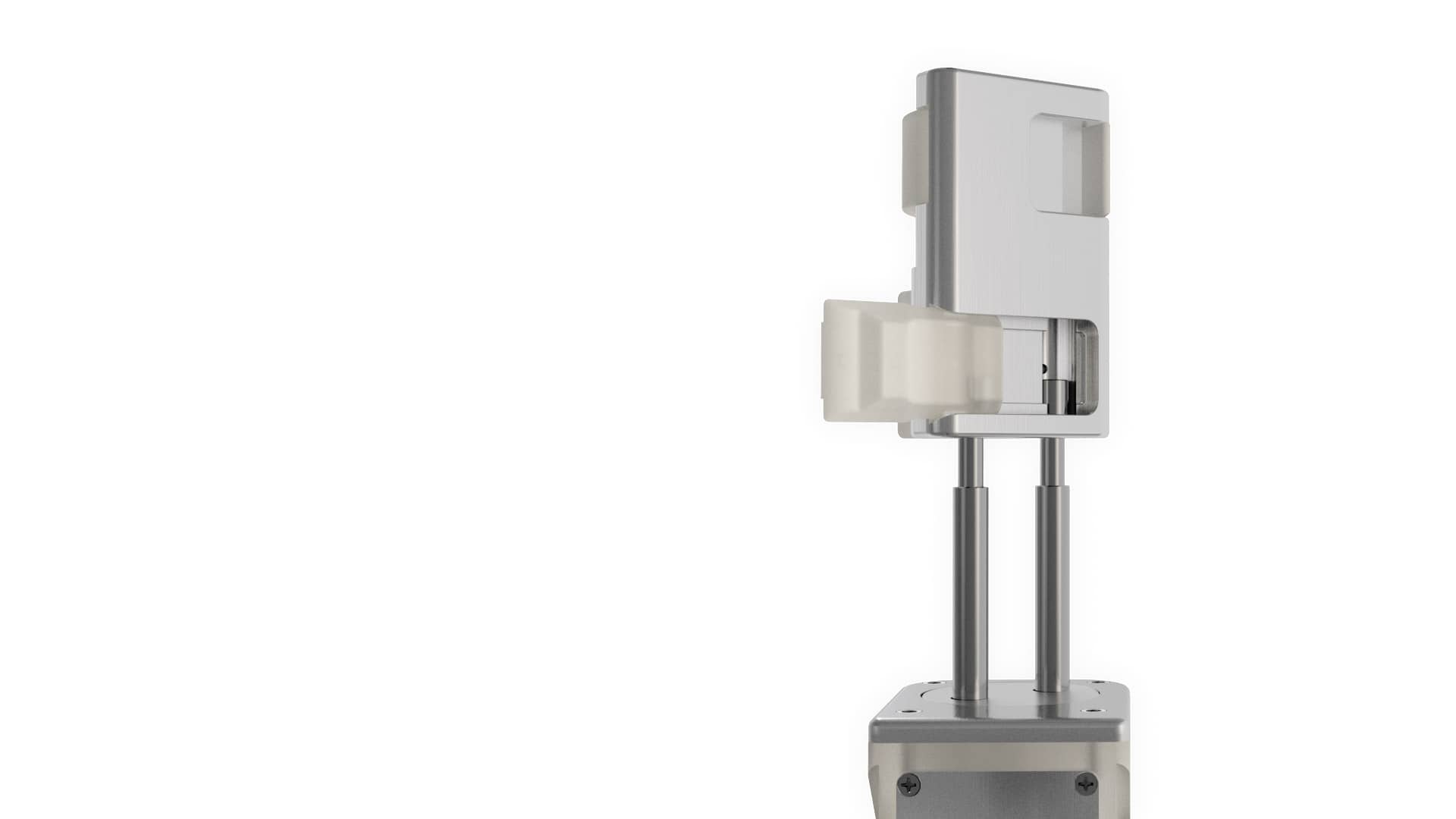
Hardware
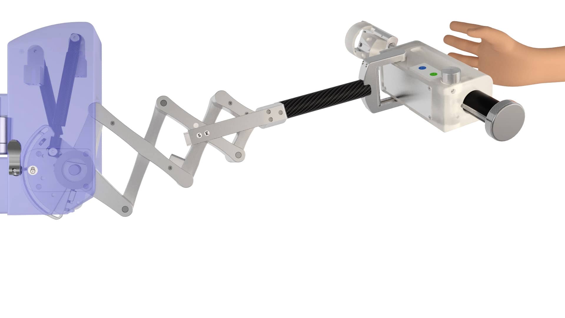
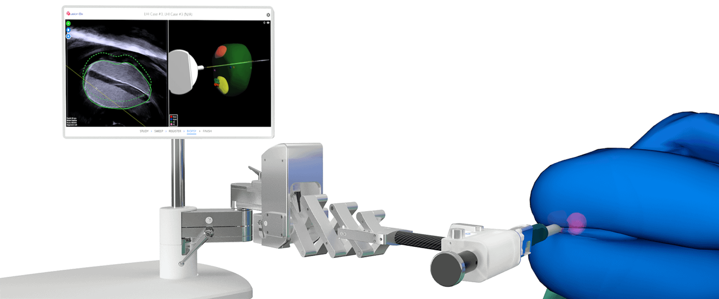
Thoughtfully engineered, the semi-robotic arm of the Fusion Bx allows you to effortlessly maneuver the probe to accurately locate and target suspicious regions of interest.
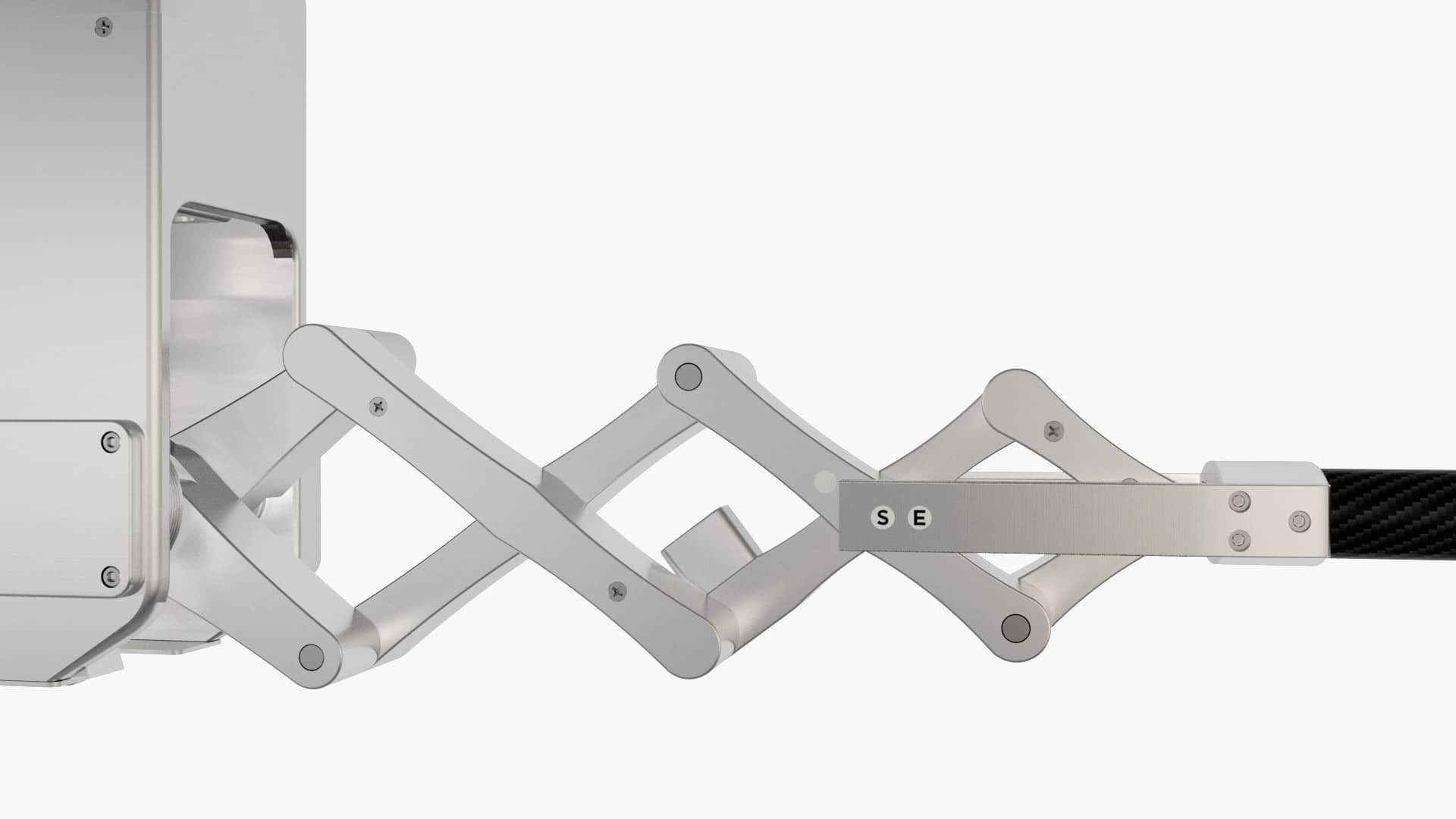
Semi-Robotic Arm
An unrestricted range of motion allows freehand-like access to the entire prostate, while maintaining consistent probe pressure to minimize prostate deformation.
Hands-Free Operation
Patented counterbalance technology simplifies procedures by supporting the ultrasound probe in any position, reducing the need for additional assistance.
Intuitive Interface
Fusion Bx is driven by a step-by-step guided workflow that enables urologists to perform procedures in less time and with minimal training. Moreover, buttons on the stepper allow urologists to advance through most of the procedure without having to take their hands off the probe.
MRI scans can be imported from USB, DVD, network or PACS.
3D model of prostate is generated from 2D ultrasound images.
Rigid and elastic registration accounts for shape and size differences.
Fused targets from MRI are superimposed onto live ultrasound.
Motion Compensation
Automatic motion compensation adjusts for patient movement to maintain image registration – allowing procedures to continue uninterrupted.
Universal Compatibility
Most ultrasound probes can be connected with ease using the corresponding holster, and both transrectal and transperineal approaches are supported.
Fusion Bx 2.0 has FDA Clearance and CE marking.
Fusion Bx 2.0 is available for sale in the United States, Canada, EU, Hong Kong, Taiwan, Singapore, Malaysia and Israel.